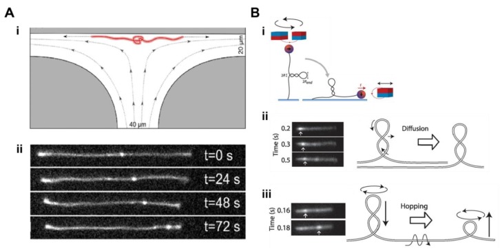
Figure 3.
Fluorescence visualisation of topological intermediates of DNA. (A) Motion of knots in DNA: (i) A microfluidic T-junction flow cell with a diverging electric field stretches knotted linear DNA molecules at its stagnation point. (ii) Representative images of a single DNA molecule at four time points as a DNA knot (bright fluorescent spot) translates towards one end of the DNA molecule (reproduced with permission from [56]). (B) Dynamics of DNA supercoils: (i) Visualisation of plectonemes by fluorescence microscopy combined with magnetic tweezers. Individual DNA molecules are supercoiled by rotating a pair of magnets and subsequently pulled sideways by another magnet. (ii) Fluorescence images of plectoneme diffusion along an individual supercoiled DNA molecule stained with SxO. (iii) Fluorescence images of a plectoneme hopping along an individual supercoiled DNA molecule stained with SxO (reproduced with permission from [66]).

