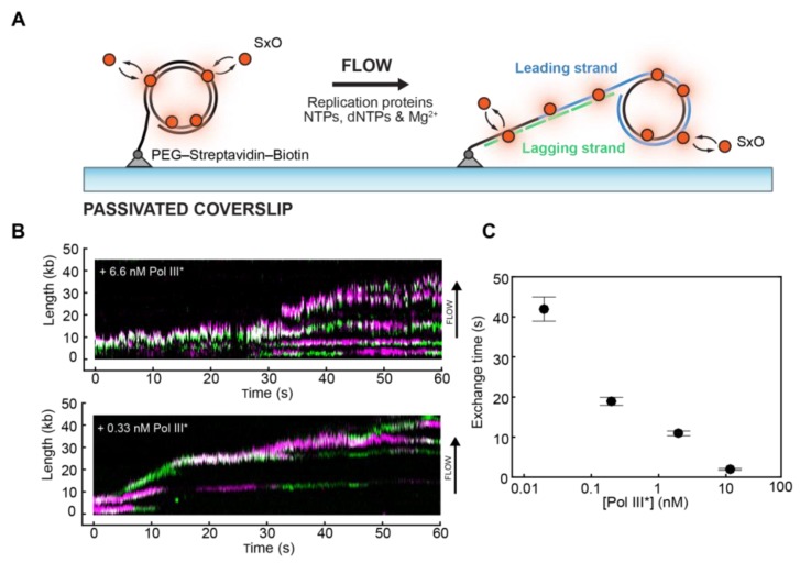
Figure 5.
Visualisation of replisome dynamics during DNA replication. (A) Cartoon representation of the single-molecule rolling-circle replication assay. A 5′-biotinylated circular DNA molecule is coupled to the surface of a passivated microfluidic flow cell through a streptavidin linkage. Addition of replication proteins and deoxyribonucleotide triphosphates (dNTPs) initiates DNA synthesis. The DNA products are elongated hydrodynamically by flow, labelled with SxO and visualised using fluorescence microscopy. (B) Rapid and frequent exchange of Pol III* (holoenzyme lacking the β2 sliding clamp) is concentration-dependent. Representative kymographs of the distributions of two different fluorescently labelled Pol III* (magenta and green) on individual DNA molecules at different concentrations. (C) Exchange times as a function of Pol III* concentration (reproduced with permission from [85]).

