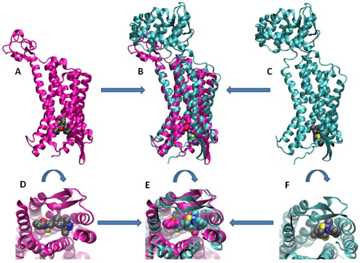
Figure 2.
Similarities in the overall structures and binding pockets of CCR5 and CXCR4. Structures of complexes are taken from the Protein Data Bank entry 6AKX (magenta) [9] and 3ODU (cyan) [10] for CCR5 and CXCR4, respectively, and superimposed by least squares fit on protein Cα atoms. (Top) Overall structure of (A) CCR5, (B) both receptors, and (C) CXCR4; (bottom) detail of the binding site of (D) CCR5, (E) both receptors, and (F) CXCR4. The ligands bound to CCR5 and CXCR4 (the 1-heteroaryl-1,3-propanediamine derivative A4R and the antagonist IT1t, respectively) are shown with atoms in standard colours (C, grey; N, blue; O, red; S, yellow) in panels A, C, D and F, and with C atoms in the same colour as their receptor (magenta for A4R, and cyan for IT1t) in panels B and E.

