Fig. 4.
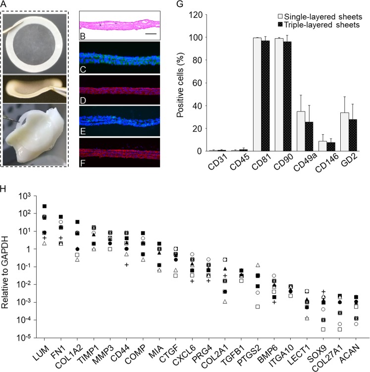
Properties of layered chondrocyte sheets are indicated. a Layered chondrocyte sheets were handled using a circular PVDF support membrane. The thin sheets attached readily to the surfaces of cartilage tissue. b–f Representative images show histological staining with hematoxylin and eosin. b Representative image showing histological staining with hematoxylin and eosin; scale bar = 100 μm. Immunohistological staining of layered chondrocyte sheets. c fibronectin, d type I collagen, e type II collagen, and f aggrecan. g Flow cytometric analysis of surface markers for chondrocytes contained in the single- and triple-layered sheets. The two kinds of cell sheets, single- and triple-layered sheets, which had been prepared for transplantation, were sacrificed and dispersed enzymatically before layering after 14 days of culture and on day 1 before transplantation, respectively. The cells sheets were analyzed by single-color staining. Data are expressed as the mean ± SD of the percentage of surface marker-expressing cells from eight independent sheets manufactured from each patient. Chondrocytes were positive for CD81 and CD90 and negative for CD31 and CD45. Staining for CD49a, CD146, and GD2 differed between patients. h The gene expression profile of transplanted sheets from each patient was analyzed using qPCR for cartilage-related genes, and the results are reported relative to the expression of GAPDH

