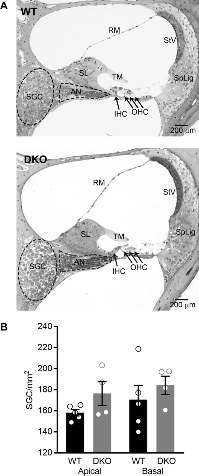
Figure 4.

Cochlear morphology and spiral ganglion cell (SGC) density are normal in KNa1 DKO mice. Cochlear morphology and SGC density were examined in mid-modiolar serial sections through the cochlea isolated from 6-week-old WT and DKO mice. (A) No differences between genotypes were seen in structures of the inner ear, including hair cells, SGCs, stria vascularis, spiral ligament and all supporting structures of the cochlear duct. (B) Spiral ganglion cell-density was not significantly different between WT (black) and DKO (grey) mice in either cochlear apical or basal turns. Data are plotted to show individual replicates (animals) and mean ± SEM. Values (mean ± SEM) and statistical analyses are provided in the Results.
