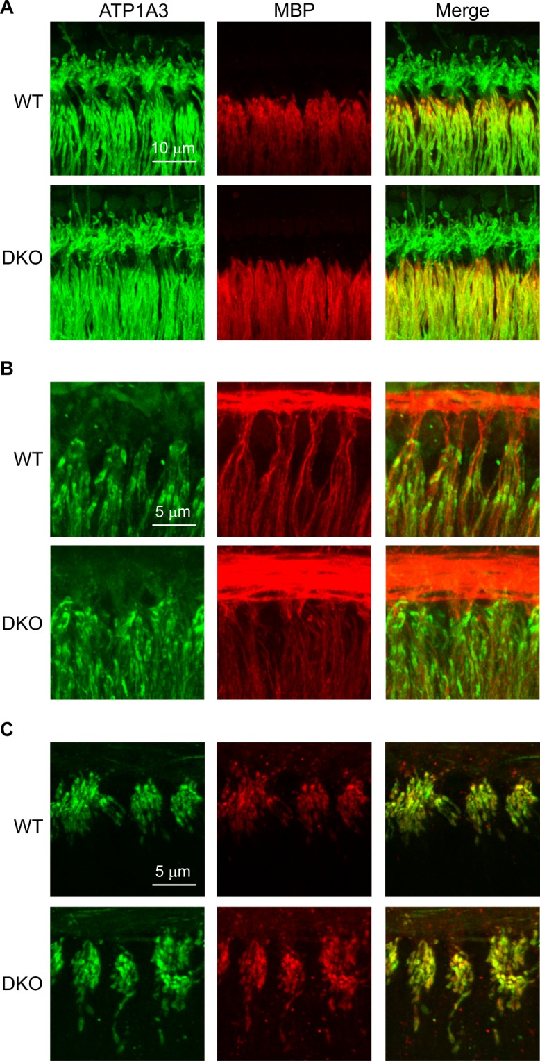
Figure 6.

Molecular and cellular architecture appears normal in SGNs from KNa1 DKO mice. The expression and distribution of various proteins shaping SGN excitability were examined using immunofluorescence in the isolated preparation of the organ of Corti and SGNs from (6-week-old) WT and DKO mice. (A) The expression of the Na+, K+-ATPase α3 (ATP1A3, green) and patterns of myelination were similar in both WT and DKO mice. (B) The expression and distribution of the low voltage-activated KV1.1 (green) was similar in both WT and DKO mice. Tubulin J (TuJ, red) marks the SGN afferent dendrites and is provided for reference. (C) The expression and distribution of the high voltage-activated KV3.1 (green) and the voltage-gated NaV1.6 (red) were similar in both WT and DKO mice. All images are presented as Z-projections through a stack of confocal micrographs from the 16 kHz region. Expression patterns of ATP1A3, MBP, KV1.1, KV3.3 and NaV1.6 were similarly expressed at other regions as well as in the somata of the SGNs from WT and DKO mice.
