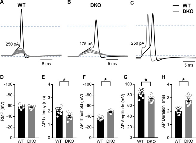
Figure 8.
Properties of evoked action potentials (APs) are altered in spiral ganglion neurons (SGNs) isolated from KNa1 DKO mice. To examine the effects of KNa1 on membrane properties in SGNs, current-clamp recordings were performed on SGNs isolated from the basal one-third of the cochlea from 6-week-old WT and DKO mice. (A) SGNs isolated from WT mice required greater current injection to evoke action potentials (APs) compared to SGNs isolated from DKO mice. (No DC current was injected to set the resting potential for different cells to the same value.) (B) Comparatively, APs evoked in SGNs from WT mice (black traces) were generally slower to initiate and larger in amplitude compared to those evoked in SGNs from DKO mice (grey traces). Dotted blue lines indicate −60 mV and 0 mV. (C–G) Across SGNs, the resting membrane potential (RMP) was not significantly different between WT and DKO mice (C). In contrast, significant differences in the AP latency, (D), membrane potential threshold for AP generation (E), AP amplitude (F) and AP duration (G) were observed between SGNs from WT and DKO mice. Data are plotted to show individual replicates (animals) and mean ± SEM. Values (mean ± SEM) from basal and apical SGNs are provided in Tables 3 and 4. For these data, significant differences between genotypes were determined using unpaired two-tailed t test.

