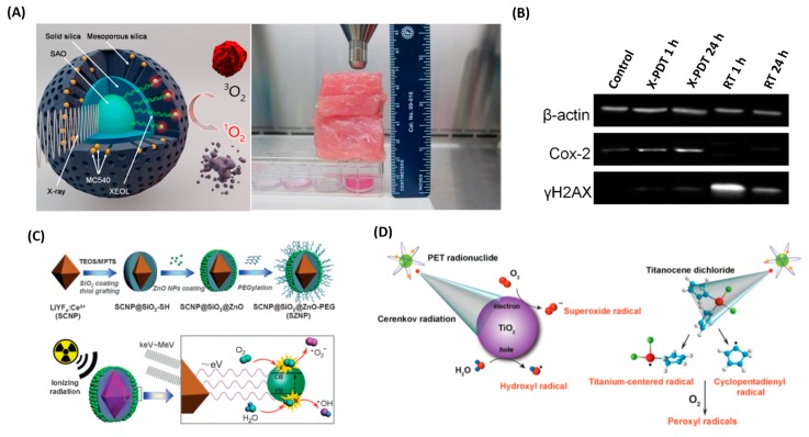
Figure 4.
(A) Schematic illustration of the working mechanism of X-PDT. Under X-ray irradiation, SAO converts X-rays to visible light photons. The visible light photons, with 4.5 cm thick pork positioned between the X-ray source and cells, activate nearby MC540 molecules to produce cytotoxic 1O2 that destroys cancer cells in the proximity. Reprinted with permission from references [77]. (B) Western blot assays, which further confirm the impact of X-PDT on DNA and membrane lipids. Reprinted with permission from references [78]. (C) Schematic illustration of the synthetic route and the mechanism of ionizing radiation-induced PDT. The electron–hole (e−–h+) pair is formed after exposure to ionizing radiation. Reprinted with permission from references [85]. (D) Schematic of CR-mediated excitation of TiO2 NP to generate cytotoxic hydroxyl and superoxide radicals from water and dissolved oxygen, respectively. Reprinted with permission from references [33].

