
Figure 2:
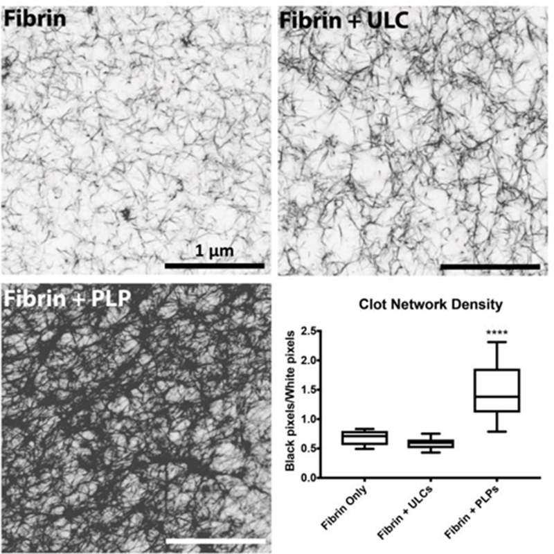
Confocal microscopy of fibrin clots taken at 63X magnification reveal that clots containing PLPs are significantly denser than control clots or clots containing ULCs, as determined by calculating black pixel area (fibrin fibers) over white pixel area (negative space). n = 3 clots/group, 3 images/clot; ****: p < 0.0001