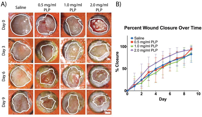
Figure 6:

PLPs enhance wound healing in vivo. A murine full-thickness dermal injury model was used to investigate dermal healing following topical application of PLPs (0, 0.5, 1.0, or 2.0 mg/ml dosages). Representative wound images over time are shown (A); outlines of the wound boundaries are shown in white. B) Percent wound closure was calculated; mean percent closure ± standard deviation are presented. n = 5 wounds/group.
