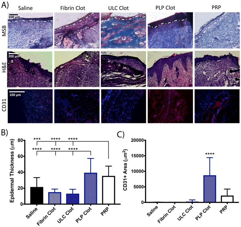
Figure 8:

A) H&E and Martius Scarlet Blue (MSB) staining of wounds harvested 9 days post-injury reveals more robust healing and epidermal layer formation in wounds treated with fibrin polymers containing PLPs relative to controls. MSB images were used to quantify epidermal thickness in (B); the outline of the epidermis on the MSB images is shown in white. Immunohistochemical analysis of wounds collected 9 days post-injury shows greater CD31+ tissue in wounds treated with fibrin polymers containing PLPs. B) Epidermal thickness, evaluated using MSB images, is significantly increased in wounds treated with fibrin polymers containing PLPs. Mean epidermal thickness ± standard deviation is shown. C) Immunohistochemical analysis of wounds collected 9 days post-injury shows increased CD31+ areas (red) in wounds treated with fibrin polymers containing PLPs. Mean CD31+ area ± standard deviation is shown. n = 5 wounds/group; ***: p < 0.001; ****: p < 0.0001.
