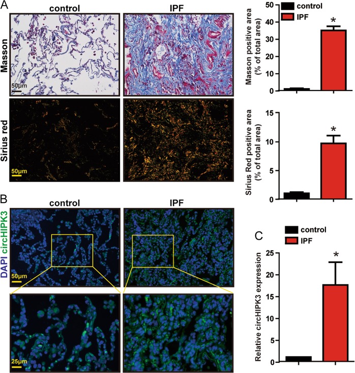
Fig. 7. Clinical relevance of circHIPK3 in idiopathic pulmonary fibrosis.
a Lung tissue sections from normal people and IPF patients were stained with Masson’s trichrome or Sirius red. Quantification of positive staining area was performed (n = 3, *p < 0.05 vs. control). Scale bar, 50 μm. b RNA-FISH assay was conducted to detect the expression distribution of circHIPK3 in lung tissues of normal people and IPF patients. Green, circHIPK3; blue, nuclei. Scale bar, upper: 50 μm; lower: 25 μm. c The expression of circHIPK3 in the lung tissues of normal people and IPF patients were detected by qRT-PCR. (n = 3, *p < 0.05 vs. control). Data are represented as means ± SD

