Figure 1.
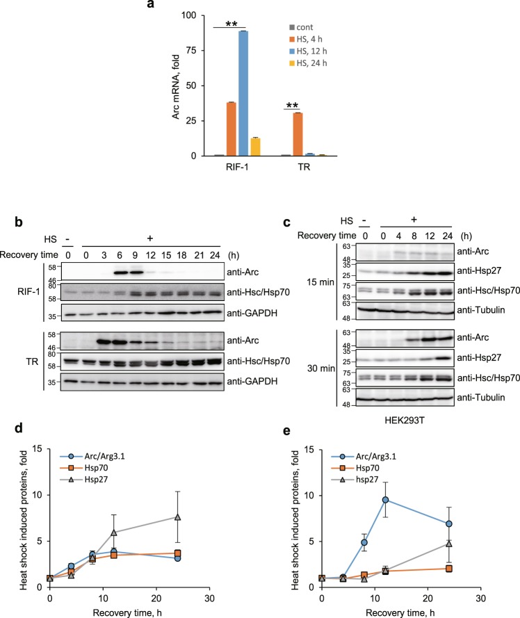
Arc/Arg3.1 is induced by heat shock. (a) RIF-1 cells were exposed to heat shock at 45°C for 30 min and recovered from the stress in fresh media at 37°C for 4, 12 and 24 h. Each sample was analyzed by Illumina microarray analysis. mRNA levels of the most up-regulated gene, Arc/Arg3.1, are shown in bar graph. Data were presented as the means ± S.D. of duplicated experiments (t-test; *P < 0.05, **P < 0.01). (b) RIF-1 and TR cells were heat shock treated at 45°C for 25 min and recovered for the indicated times in fresh media. Arc/Arg3.1 and Hsc/Hsp70 were analyzed by Western analysis with their specific antibodies. As a loading control, GAPDH level was detected using anti-GAPDH antibody. Western blot results were selected representative data from triplicated results. (c) HEK293T cells were exposed to heat shock at 45°C for 15 and 30 min and recovered for the indicated times. Arc/Arg3.1, Hsp27 and Hsc/Hsp70 were detected by Western blot analysis with their specific antibodies. As a loading control, tubulin level was detected using anti-tubulin antibody. Western blot results were selected representative data from triplicated results. Quantified results of triplicated Western images were shown in (d,e) for 15 min and 30 min heat shock treatment, respectively. Data were presented as the means ± S.D.

