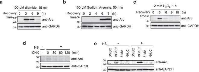
Figure 2.
Arc/Arg3.1 is induced by various stresses and degraded in proteasome. (a–c) HeLa cells were treated with 100 μM diamide for 15 min (a), 100 μM sodium arsenite for 50 min (b) or 2 mM H2O2 for 1 h (c). Cells were recovered for the indicated times with fresh media. Arc/Arg3.1 was analyzed by Western analysis using anti-Arc/Arg3.1 antibody. As loading controls, GAPDH and tubulin levels were detected using anti-GAPDH and anti-tubulin antibody. (d) HeLa cells were heat shock treated at 45°C for 25 min and recovered for 9 h in fresh media at 37°C to induce maximum Arc/Arg3.1 expression. Then cells were treated with 10 μg/mL cycloheximide (CHX) for the indicated times. The endogenous Arc/Arg3.1 was detected by Western analysis using anti-Arc antibody. As a loading control, GAPDH level was detected using anti-GAPDH antibody. (e) After heat shock stress at 45°C for 25 min, HeLa cells were recovered for 9 h in fresh media. Then cells were incubated with 10 μM MG132, 10 mM 3-methyladenine (3-MA) or 10 mM NH4Cl for 6 h. The cell were analyzed by Western analysis using anti-Arc/Arg3.1 antibody. As a loading control, GAPDH level was detected using anti-GAPDH antibody. Western blot results were selected representative data from more than triplicated results.

