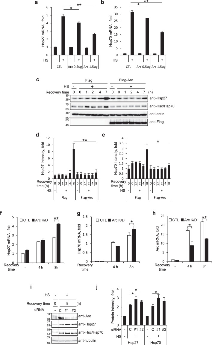
Figure 3.
Arc/Arg3.1 inhibits Hsp27 and Hsp70 induction. (a,b) HEK293T cells were transfected with Flag empty vector or Flag-Arc. Cells were exposed to heat shock at 45°C for 15 min and recovered for 5 h. Quantitative RT-PCR was performed using primers for Hsp27 (a) and Hsp70 (b) mRNA. Relative quantities of Hsp27 mRNA and Hsp70 mRNA were normalized against GAPDH mRNA. (c–e) HEK293T cells overexpressing Flag or Flag-Arc/Arg3.1 were exposed to heat shock at 45°C for 15 min and recovered for the indicated times. Cells were analyzed by Western blot analysis using anti-Hsp27, anti-Hsp70, anti-β-actin and anti-Flag antibodies (c). Quantified amounts of Hsp27 (d) and Hsp70 (e) were represented. (f–h) HeLa cells were transfected with control siRNA and Arc/Arg3.1 siRNA #1. After 48 h, cells were exposed to heat shock at 45°C for 15 min, and recovered at 37°C for the indicated times. HSP27 mRNA (f), HSP 70 mRNA (g) and Arc/Arg3.1 mRNA (h) were analyzed using quantitative RT-PCR and normalized to GAPDH mRNA. (i,j) HEK293T cells were transfected with control siRNA and Arc/Arg3.1 siRNAs (#1, #2), heat shock treated at 45°C for 15 min and recovered for 4 h or 8 h. Cells were analyzed by Western blot analysis using anti-Arc, anti-Hsp27, anti-Hsp70 and anti-tubulin antibodies (i). Quantified amounts of Hsp27 and Hsp70 in 8 h recovered cells were represented (j). Data were presented as the means ± S.D. of triplicated experiments (t-test; *P < 0.05, **P < 0.01). Western blot results were selected representative data from more than biologically triplicated results.

