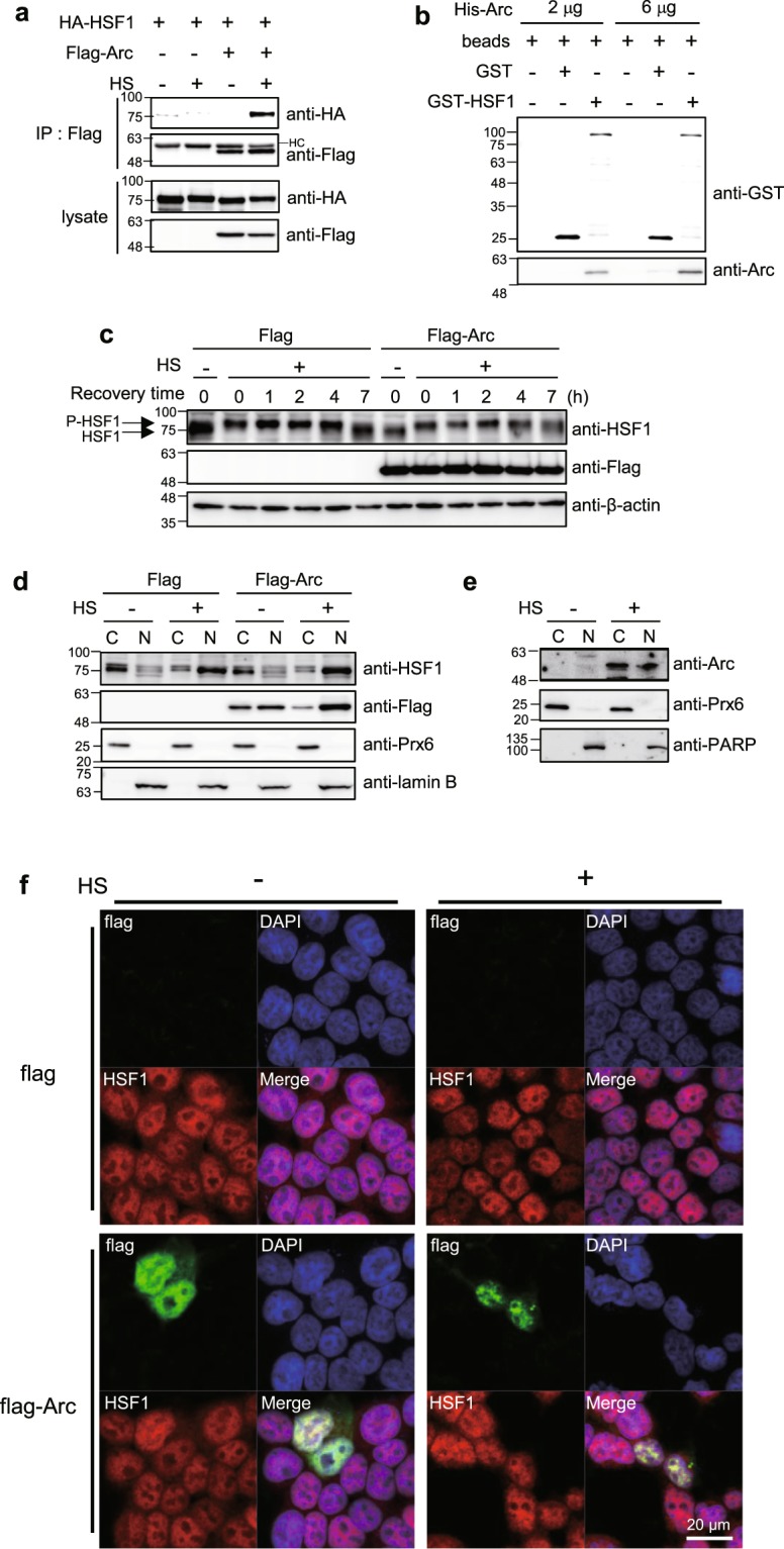
Figure 4.

Arc/Arg3.1 binds with HSF1. (a) HEK293T cells were transfected with HA-HSF1 and Flag empty vector or Flag-Arc. After exposure to heat shock at 45°C for 15 min, immunoprecipitation was performed using anti-Flag antibody. Protein complex was analyzed by Western blot analysis using anti-HA and anti-Flag antibodies. (b) GSH beads, beads bound GST and beads bound GST-HSF1 were incubated with purified His-Arc. After washing step, beads bound proteins were analyzed by Western analysis using anti-GST and anti-Arc antibodies. (c) HEK293T cells were transfected with Flag empty vector or Flag-Arc. Cells were exposed to heat shock at 45°C for 15 min and recovered for the indicated times. Cells were analyzed by Western analysis using anti-HSF1, anti-Flag and anti-β-actin antibodies. P-HSF1; phosphorylated HSF1. (d) HEK293T cells were transfected with Flag empty vector or Flag-Arc. Cells were exposed to heat shock at 45°C for 15 min and fractionated into cytosolic (C) and nuclear (N) fractions. Each fraction was analyzed by Westen analysis using anti-HSF1, anti-Flag, anti-Prx6 and anti-Lamin B antibodies. Prx6; the cytosol marker, lamin B; the nucleus marker. (e) Hela cells were heat shock treated at 45°C for 25 min and fractionated into cytosolic (C) and nuclear (N) fractions. Each fraction was analyzed by Western analysis using anti-Arc, anti-Prx6 and anti-PARP antibodies. Prx6; the cytosol marker, PARP; the nucleus marker. (f) HEK293T cells were plated on the glass coverslip 24 h before transfection. Cells were then transfected with Flag or Flag-Arc. After 24 h, cells were treated with heat shock at 45°C for 15 min and visualized Flag-Arc (green), HSF1 (red) and nucleus (blue) under confocal microscopy. All of the Western blot results were selected representative data from more than duplicated results.
