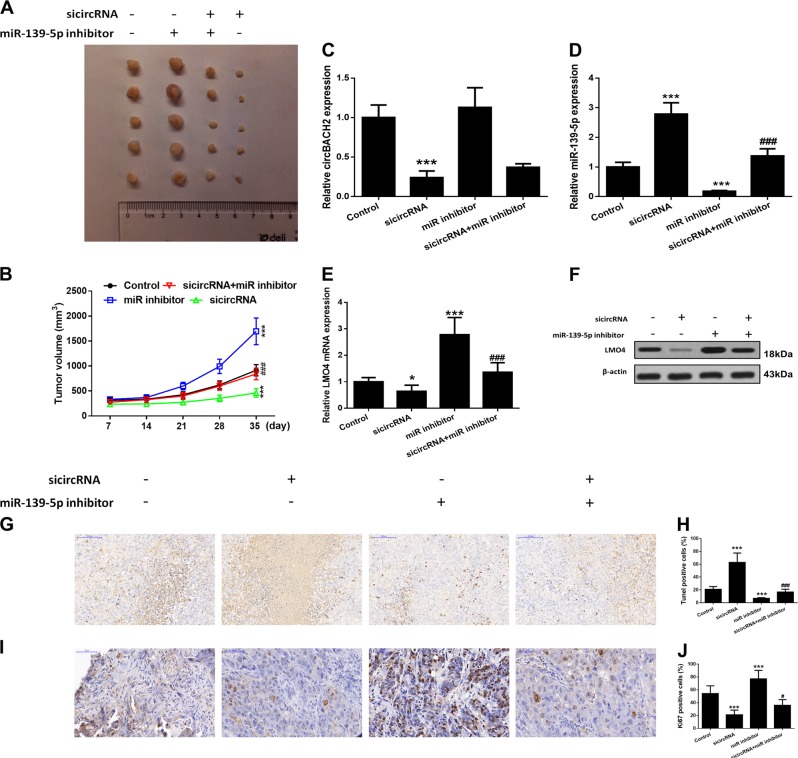
Fig. 6. Effects of circBACH2 or miR-139-5p in a tumor xenograft model.
TPC-1 cells stably inhibiting circBACH2 or miR-139-5p were inoculated subcutaneously into the right flank regions of 4-week-old male BALB/c nude mice. a Representative images of xenograft tumors isolated from nude mice in the different groups. b Tumor sizes in the different groups. c, d The expression of circBACH2 and miR-139-5p was detected by RT-PCR in the indicated groups of tumors. e, f The expression of LMO4 was detected by RT-PCR and western blotting in the indicated groups of tumors. g, h The TUNEL assay (400×) was performed to determine the apoptotic indices and quantified. i, j The Ki-67 assay (400×) was performed by immunohistochemistry and quantified. Scale bar, 100 μm. Data indicate the mean±SD, n = 5. **P < 0.01, ***P < 0.001 vs. control and ###P < 0.001 vs. sicircRNA

