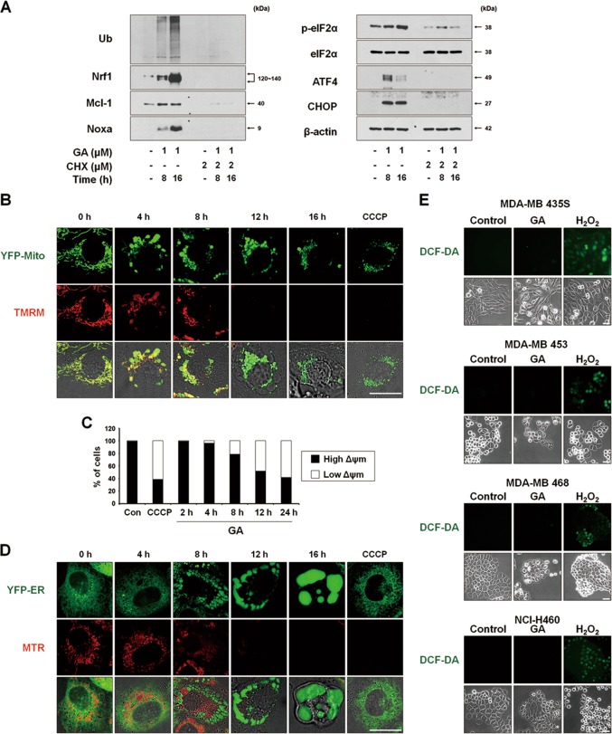
Fig. 4. ROS levels are not noticeably increased in spite of the MMP loss in GA-induced paraptosis.
a MDA-MB 435S cells pretreated with or without 2 μM CHX and further treated with 1 μM GA for the indicated time points. Cell extracts were prepared for Western blotting of the indicated proteins. Western blotting of β-actin was used as a loading control. b, c YFP-Mito cells (b) or MDA-MB 435S cells (c) were treated with 1 μM GA for the indicated time points or 20 μM CCCP for 12 h were incubated with TMRM. Samples were subjected for confocal microscopy (b) or flow cytometry (c). d YFP-ER cells were treated with 1 μM GA for the indicated time points or 20 μM CCCP for 12 h. Treated cells were incubated with MTR and observed under the confocal microscope. e Cells treated with GA (1 μM for MDA-MB 435S; 2 μM for MDA-MB 453; 3 μM for MDA-MB 468 and NCI-H460 cells) for 16 h or 5 mM H2O2 for 10 min were incubated with CM-H2DCF-DA (DCF-DA) and subjected for the fluorescence microscopy. Bars, 40 μm

