
Figure 2.
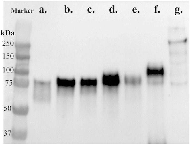
Western blotting of HEK293 cells expressing various truncated spike proteins. Cell lysates of HEK293 cells expressing different lengths of S protein were separated by SDS-PAGE, transferred to PVDF membranes, probed with an anti-V5 tag antibody, and detected using HRP-conjugated secondary goat anti-mouse immunoglobulin antibody. The protein ladder/marker is shown in kilodalton (kDa). The bands corresponding to the various truncations of the spike protein are (a) a.a. 1–435; (b) a.a. 1–485; (c) a.a. 1–501; (d) a.a. 1–509; (e) a.a. 1–575; (f) a.a. 1–639; (g) full-length spike protein.