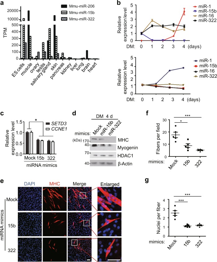
Fig. 4. Overexpression of miR-15b or miR-322 delayed myoblast differentiation.
a The expression levels of the indicated miRNAs in embryonic stem (ES) cells and various mouse tissues were plotted using the datasets in GEO. b The expression levels of the indicated miRNAs during C2C12 cells differentiation were analysed by RT-qPCR (top panel) or by using the datasets in GEO (bottom panel). c–g C2C12 cells were transfected with the indicated miRNA mimics, and induced differentiation for 4 days. c The mRNA levels of SETD3 and CCNE1 from differentiated cells were examined by RT-qPCR. d The differentiation protein markers in the transfected cells were analyzed by western blot analysis. e Immunofluorescence staining was performed using a specific antibody against MHC (red). DAPI stains nuclei. Scale bar: 100 μm. f MHC-positive cell numbers in certain field of the indicated C2C12 cells describe in e were quantified. g Quantitative analyses of nuclei number per fiber from the indicated C2C12 cells. Data represent the mean ± SD from three independent experiments

