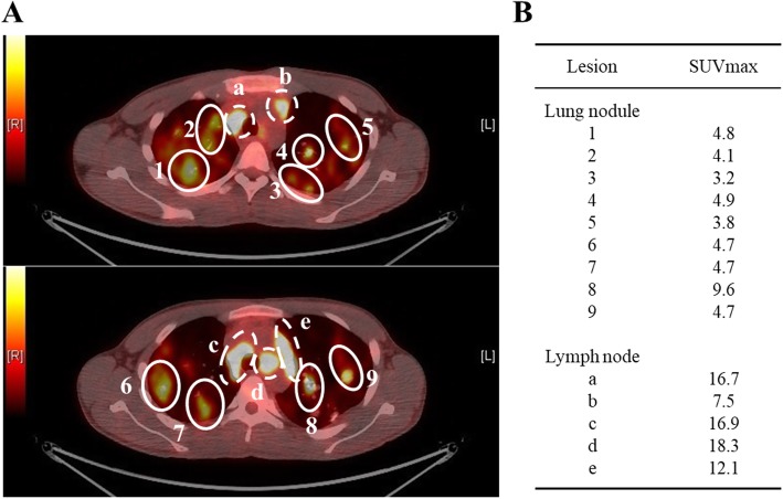
Fig. 2.
The axial view of FDG-PET/CT scan on diagnosis. (A) PET/CT scan shows intense FDG uptake in bilateral lung masses and the mediastinal and hilar lymph nodes. Each lung nodule is surrounded by a solid-line circle (1–9). Each mediastinal lymph node is surrounded by a dotted line circle (a-e). (B) The maximum standardized uptake values (SUVmax) of the lesions are shown here

