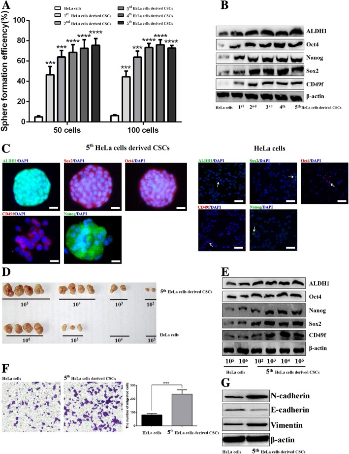
Fig. 1.
Resuscitated HeLa cells derived CSCs show stemness phenotypic characteristics. The graph shows the SFE of 1st to 5th- passaged HeLa cells derived CSCs and parental HeLa cells (a). Western blot analysis of ALDH1, Sox2, CD49f, Nanog, and Oct4 in 1st to 5th-passage HeLa cells derived CSCs and parental HeLa cells (b). Immunofluorescence staining of ALDH1, Sox2, CD49f, Nanog, and Oct4 in 5th-passage HeLa cells derived CSCs and parental HeLa cells, respectively; the white arrows point to positive cells (c). Injection of different density of 5th-passage HeLa cells derived CSCs and parental HeLa cells generated xenografts in nude mice (d). Western blot analysis of ALDH1, Sox2, CD49f, Nanog, and Oct4 in tumor tissues derived from 5th-passage HeLa cells derived CSCs or HeLa cells bearing mice (e). Transwell assay showing the migrated cells of 5th-passage HeLa cells derived CSCs and parental HeLa cells; the histogram shows the number of migrated cells; original magnification, × 400 (f). Western blot analysis of E-cadherin, Vimentin, and N-cadherin in 5th-passage HeLa cells derived CSCs and parental HeLa cells (g). * P < 0.05, ** P < 0.01, *** P < 0.001. Scale bars represent 50 μM or 10 μM in inset. Results are shown as mean values ± SD of independent experiments performed in triplicate

