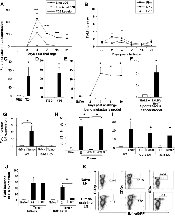
Figure 1.
Increased IL4 expression in tumor-draining lymph nodes (LN). A and B, 2.5 × 10⁵ live or irradiated CT26 colon cancer cells or CT26 cell lysate were injected subcutaneously. Tumor-draining lymph nodes were removed as indicated and analyzed for IL4, IFNγ, IL10, and IL13 mRNA expression. C and D, 2.5 × 10⁵ TC-1 cervical or 4T1 breast cancer cells were injected subcutaneously into the right flank of syngeneic mice and IL4 expression examined 7 days later. E, 2.5 × 10⁵ 4T1 cells were injected intravenously and IL4 mRNA levels examined over 16 days. F, Tumor-draining lymph nodes from 4-month-old BALB-neuT mice were removed and analyzed for IL4 mRNA. A total of 2.5 × 10⁵ MC38 (G, I) or CT26 (H, J, K) colon cancer cells were injected subcutaneously into C57BL/6, RAG1 KO, BALB/c 4GET, CD1d KO, Jα18 KO, or CD11c-DTR/EGFP mice. H, BALB/c mice were also injected intraperitoneally with 500 μg of depleting antibodies to CD4 or CD8 on day 1. Tumor-draining lymph nodes were removed on day 7 and analyzed for IL4 mRNA levels (G–J). J, CT26 colon cancer cells were injected subcutaneously into CD11-DTR or BALB/c mice that were injected the next day with 100 ng of diphtheria toxin (DT). Tumor-draining lymph nodes were removed and analyzed for IL4 mRNA (J). All results represent the mean + SD of results from 4 to 8 independent lymph nodes. Experiments were repeated two or three times with similar results. K, Naïve or tumor-draining lymph nodes from 4GET mice were removed on day 7 and analyzed for the expression of eGFP and TCRβ, CD3ε, or CD4 by flow cytometry. *, P < 0.01 when compared with PBS-treated or naïve group. **, P < 0.01 when compared with irradiated tumor cell–treated group.

