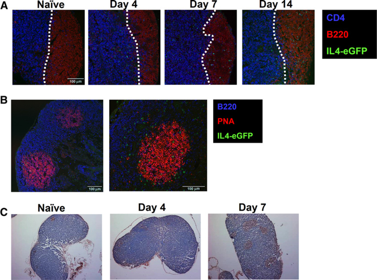
Figure 4.
IL4-expressing cells localized to the germinal center of tumor-draining lymph nodes (LN). Confocal imaging of tumor-draining lymph nodes from 4GET mice. A, Time course analysis showing CD4 (blue), B220 (red) and IL4-eGFP (green). B, Analysis on day 14 showing B220 (blue), PNA (red), and IL4-eGFP (green). C, Section of tumor-draining lymph nodes at indicated days stained with PNA (brown). Experiments were repeated five times with similar results.

