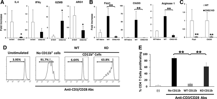
Figure 6.
Altered gene expression and suppressive function of myeloid cells in CNS2 KO mice. A, 105 CT26 cells were injected subcutaneously into WT/CNS2 HT or CNS2 KO mice. CT26 tumors were removed at day 21 and analyzed for indicated mRNA by real time qPCR. B, CD11b+ myeloid cells from lymph nodes (LN) of CT26 tumor bearing mice were isolated by MACS and analyzed for Fizz1, Chi3l3, and ARG-1 mRNA by real time qPCR. Mean SD, from 6 to 8 independently analyzed mice per group, are shown. C, Tumors were removed on day 25 and sorted by FACS based on their expression of CD45+, CD11b+, Ly6c−, and Ly6g−. Sorted macrophages were analyzed for mRNA expression. D and E, CD11b+ cells from the spleen of CT26 tumor-bearing WT or CNS2 KO mice were isolated on day 25 post-tumor challenge by MACS (final purity, 90%–95%). A total of 5 × 10⁵ cells were cultured for 4 days with 2.5 × 10⁵ CFSE-labeled syngeneic CD4 T cells in the presence or absence of 0.1 mg/mL anti-CD3/anti-CD28. T-cell proliferation was monitored by CFSE dilution. D, Representative example and (E) mean SD (n = 4 independent CD11b+ cells preparations in three independent experiments) are shown; *, P < 0.05. **, P < 0.01.

