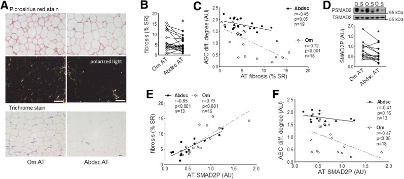
Figure 7.
Higher pericellular fibrosis in Om was correlated positively with tissue TGFβ signaling activity and negatively with ASC differentiation (diff.). A: Adipose tissue (AT) samples were used for picrosirius red and trichrome staining of collagen, and representative images from a donor are presented. Scale bars, 100 µm. B: Levels of pericellular fibrosis were quantified in picrosirius red–stained slides and presented as percent of stained to total area (% SR). *P < 0.05 (paired t test), n = 20 (mean ± SEM BMI 42.7 ± 2.1 kg/m2 [range 23–63] and age 38.7 ± 2.6 years [20–56], with 14 females and 6 males). C: Inverse correlations between pericellular fibrosis and differentiation degree of ASCs derived from the same donors. D: SMAD2P in Om and Abdsc adipose tissues was measured with immunoblotting. *P < 0.05 (paired t test; n = 14). E: Positive associations between tissue SMAD2P and fibrosis (percent of stained to total area). F: Negative associations between tissue SMAD2P and differentiation degree of ASCs derived from the same donors. The numbers of subjects, Pearson correlation coefficients (r), P values, and number of samples are shown in each panel. AU, arbitrary units. O, Om; S, Abdsc.

