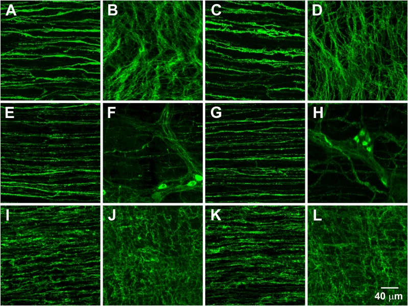
Figure 3.
Networks of ICC, NOS1+ nerves, and PDGFRα+ cells along the greater curvature of antrums from wild-type and Lepob animals. Representative images of ICC (A–D), NOS1+ neurons (E–H), and PDGFRα+ cells (I–L). A, E, and I show ICC, NOS1+ nerve fibers, and PDGFRα+ cells, respectively, within the circular muscle layer of a wild-type antrum. B, F, and J show ICC, NOS1+ neurons, and PDGFRα+ cells, respectively, in the myenteric plexus region of a wild-type antrum. C, G, and K show ICC, NOS1+ nerve fibers, and PDGFRα+ cells, respectively, within the circular muscle layer of a Lepob antrum. D, H, and L show ICC, NOS1+ neurons, and PDGFRα+ cells, respectively, in the myenteric plexus region of a Lepob antrum. Scale bar in L applies to all panels.

