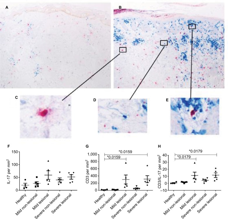
Figure 2.
Marked increases in IL-17 expression and CD3+ T cells within the dermis of affected skin.
Notes: (A) Immunostaining of IL-17 (red) and CD3 (blue) of an uninvolved skin biopsy of a psoriatic patient. (B) Immunostaining of IL-17 (red) and CD3 (blue) of a skin biopsy out of a psoriatic plaque (lesional skin). (C) Detailed view of IL-17 staining in immunohistochemical analysis. (D) Detailed view of a CD3+ cell in the immunostaining. (E) Detailed view of a IL-17+ CD3+ cell in the double immunostaining. (F–H) IL-17, CD3, and IL-17+ CD3 double staining in the dermis of healthy controls (n=4) (F) and patients with mild (n=5) (G) and severe (n=5) (H) psoriasis vulgaris assessed with immunostaining. Cutaneous IL-17 expressing cells/mm2 in individual patients as well as the arithmetic mean are shown. *P<0.05, Mann–Whitney U test.

