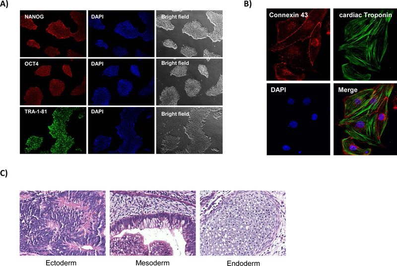
Figure 3.

Immunofluorescence staining demonstrating (A) pluripotency markers NANOG, OCT4 and TRA-1–81, DAPI, and bright field images. (B) Immunofluorescent staining of iPSC-derived cardiomyocytes demonstrating positive staining of cardiac markers connexin 43 and cardiac troponin as well as DAPI. (C) Representative H&E staining from teratomas to confirm the pluripotency status of the generated iPSCs.
