
Figure 3: LC-NA and LC-GABA neurons receive inputs from similar as well as different sources.
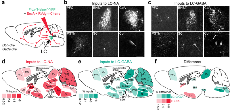
a. Schematic for targeting pseudo-rabies virus to LC-NA and LC-GABA subpopulation. b,c. Transynaptically labelled neurons in different brain regions following injection of targeted pseudo-rabies virus in LC of Dbh-Cre or Gad2-Cre mice. Repeated in N = 8 and 4 mice for LC-NA and GABA respectively. Scale bars: 200 μm. d,e. Map of brain regions providing the largest fraction of inputs to LC-NA and LC-GABA neurons. Regions providing less than 0.5% of total inputs are not displayed. N = 8 and 4 mice for LC-NA and LC-GABA respectively. f. Map of the difference in inputs to LC-NA and LC-GABA neurons. Only regions showing significant difference are displayed (p < 0.05 using paired t-test; see Supplementary Table 1). BNST: bed nucleus of the stria terminalis; Cb: cerebellum; CbN : cerebellar nuclei; CeA: central amygdala; CnF: cuneiform nucleus; DB: diagonal band; DpMe: deep mesencephalic nucleus; Gi: gigantocellular nucleus; IC: inferior colliculus; IPL: interpeduncular nucleus; LH: lateral hypothalamus; LHb: lateral habenular nucleus; MC: motor cortex; PAG: periaqueductal gray; PaV: paraventricular nucleus; PFC: prefrontal cortex; PH: posterior hypothalamus; PnO: pontine nucleus; PO: preoptic nucleus; Pr: prepositus nucleus; PSTh: parasubthalamic nucleus; Rt: reticular nucleus; SC: superior colliculus; SN: substantia nigra; Sp5: spinal trigeminal tract; SPF subparafascicular thalamic nucleus; SuM: supramammillary nucleus; ZI: zona incerta.