Figure 13.

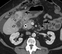
Gallbladder carcinoma. CT of a patient with biopsy‐proven gallbladder carcinoma demonstrates loss of the fat plane between the gallbladder and adjacent liver, and a thickened gallbladder wall with calcifications (black arrows).

Gallbladder carcinoma. CT of a patient with biopsy‐proven gallbladder carcinoma demonstrates loss of the fat plane between the gallbladder and adjacent liver, and a thickened gallbladder wall with calcifications (black arrows).