Figure 3.
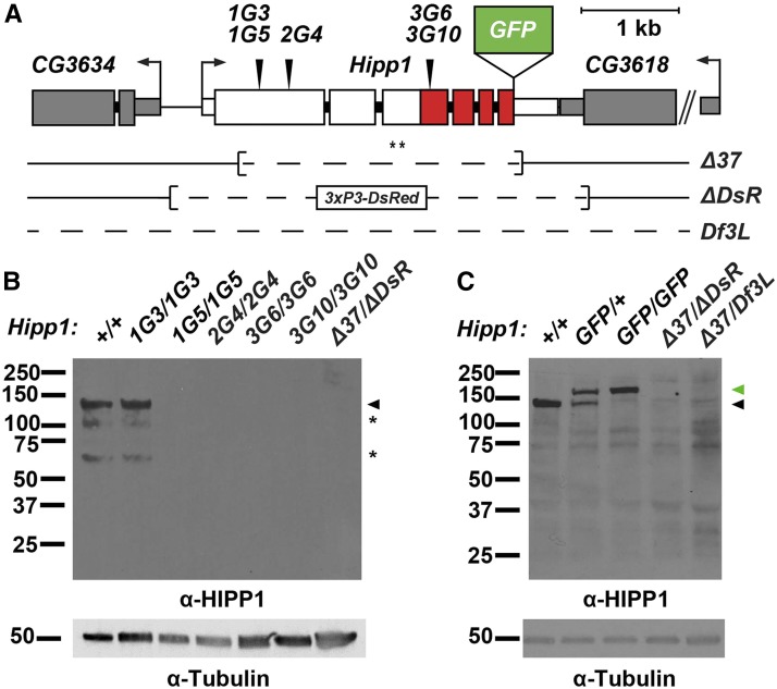
Structure of the Hipp1 locus. A. Shown is the structure of the Hipp1 gene, with exons (large rectangles) colored to indicate the positions of the crotonase-like domain (CLD, red) and bent arrows to show directions of transcription. Structures of the neighboring CG3634 and CG3618 genes are indicated in gray. Inverted triangles above the Hipp1 gene indicate the locations of the small CRISPR generated deletions (1G3, 1G5, 2G4, 3G6 and 3G10), whereas the locations of the large deletions (Δ37 and Δ DsR) are shown below the gene. The position of insertion of the GFP coding region is shown (raised green rectangle). Asterisks indicate the location of the peptide epitopes recognized by the HIPP1 antibody. B., C. Western blot of protein extracts obtained from ovaries dissected from wild type (+/+, Canton S) or Hipp1 mutant females of the indicated genotype. Blots were probed with the HIPP1 antibodies, using antibodies against alpha-Tubulin as a loading control. Positions of full-length proteins are shown by black and green arrowheads, indicating HIPP1 and HIPP1-GFP, respectively. Asterisks indicate positions of degradation products.

