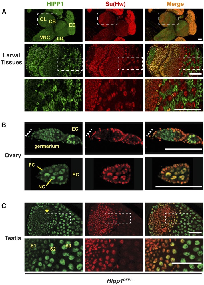
Figure 4.
HIPP1 is a globally expressed nuclear protein. A-C. Confocal images of tissues dissected from Hipp1GFP/+ and stained with antibodies against GFP (HIPP1, green) and Su(Hw) (red), with the merged image at the right. A. Top panels: Representative images of tissues dissected from third instar larvae, showing neuronal tissues of the central brain (CB), optic lobe (OL), ventral nerve cord (VNC), as well as non-neuronal tissues (eye disc, ED; leg disc, LD). Bottom panels: Magnification of boxed region of the central brain isolated from a Hipp1GFP/+ wandering third instar larva. This section reveals that some cell types express Su(Hw), but not HIPP1. Scale bars, 50 μm. B. Top panels: Image of a germarium, with the position of the somatic niche shown as a dashed line. Bottom panels: an early stage egg chamber (EC, bottom) that contains differentiated germ cells (nurse cells, NC) surrounded by somatic follicle cells (FCs). Scale bars, 20 µm. C. Top panels: Image of a testis that shows the somatic niche (hub, asterisk) and developing germ cell cysts. Bottom panels: Magnification of the boxed region to highlight the transition between Su(Hw) positive spermatocytes (stages S1 to S2) and Su(Hw) absent spermatocytes (S3). HIPP1 expression is stronger in mid-to-late stage spermatocytes. Scale bars, 50 µm.

