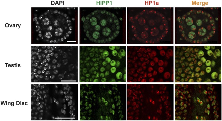
Figure 5.
HIPP1 and HP1a show limited co-localization in interphase cells. A. Top: Representative confocal image of an early stage egg chamber (stage 4) in an ovary dissected from 1< day-old Hipp1GFP/+ female stained with DAPI, α-GFP (green), and α-HP1a (red). Middle: Representative confocal image of the anterior portion of a 1< day-old testis dissected from a Hipp1GFP/+ male and stained as described in A. Scale bars, 20 μm. Anterior is to the left. In testes, HP1a localizes diffusely in spermatocyte nuclei. Bottom: Representative confocal image of a third instar larval wing disc stained as described in A.

