Figure 3.
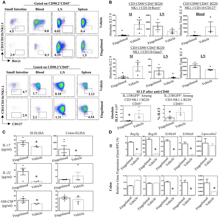
Fingolimod causes ILC-penia, augments lymph node ILC numbers, and decreases small intestine lamina propria ILC3 numbers in mice but does not reduce antimicrobial peptide production. (A) A representative flow plot of the percentages of total ILCs in the small intestine lamina propria blood, inguinal lymph node (LN) and spleen of mice gavage-fed with fingolimod or vehicle for 30 days. The plots show live cells gated on CD90.2+CD45+B220- which then plotted as CD3/NK1.1/CD11b vs. Rorγt (top panel) or Gata3 (bottom panel). (B) Absolute number of Rorγt+ ILC3s (top panel), of Gata3+ ILC2s (middle panel) in the steady state mice after fingolimod of vehicle-gavage feeding for 30 days. Top panel, right, shows total ILCs (CD90.2-CD45+CD3-B220-CD11b-NK1.1-CD127+) in the blood of steady state mice following 30 days of fingolimod or vehicle treatment. The bottom most panel shows absolute number or percentages of IL-23RGFP+ ILC3s in the small intestine lamina propria 2-days after anti-CD40 injection following 30 days fingolimod or vehicle treatment. Three-to-four mice per group were used. Experiment was repeated 3 times. (C) 1 cm piece of ileum from fingolimod or vehicle-injected mice (for 30 days) were cultured 48 h and the supernatants were assessed with ELISA for the production of indicated ILC3-associated cytokines. Four to five of mice per group. (D) 1 cm piece of ileum or colon from fingolimod or vehicle-injected mice for 30 days was examined for gene expression of indicated antimicrobial peptides via real-time qPCR. *Indicates p < 0.05.

