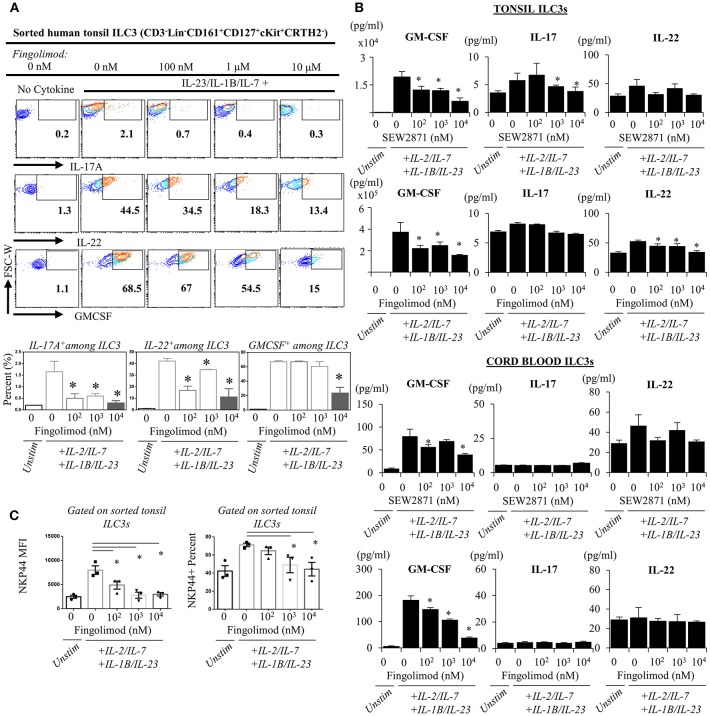
Figure 4.
Ex vivo exposure of human ILC3s to S1P analogs inhibits production of ILC3-associated cytokines. (A) Sorted ILC3s from tonsils (gated as Lin-CD3-CD161+CD127+ckit+ CRTH2-) were cultured in complete medium at increasing doses of FTY720 and activated with IL-2, IL-23, IL-7, and IL-1B (20 ng/ml each) for 2–3 days, and PMA (50 ng/ml) /Inonomycin (1 μg/ml) stimulated for 4 h at 37°C for intracellular staining of indicated cytokines, representative flow plots (left) and quantified bar graphs for percentages of cells producing the indicated cytokines (right). (B) ELISA was performed from supernatants of “A” and SEW2871 exposed tonsil-derived (top panel) cord blood-derived (bottom panel) ILC3 cultures for ILC3-asssociated cytokines cord-derived ILC3s. (C) NKP44 surface expression by ILC3s were examined via flow cytometry, percent and mean fluorescence intensity (MFI) was quantified after culture with FTY720 for 3 days. *Indicates p < 0.05. The experiments were performed with triplicates and repeated for 3 times.

