Figure 3.

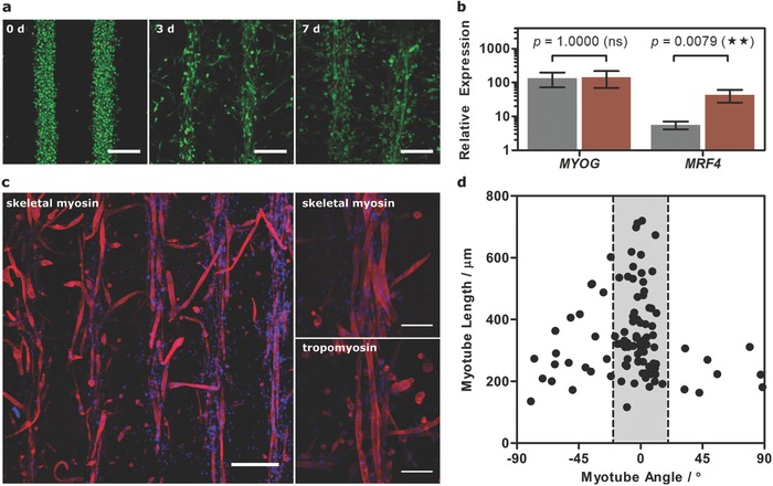
Engineering patterned muscle using GelMA. a) Confocal fluorescence microscopy of acoustically patterned myoblasts in 40 mg mL−1 GelMA over 7 d of tissue engineering. Myoblasts were stained with calcein (green, viable cells) and ethidium homodimer (red, nonviable cells). Scale bars, 200 µm. b) Relative expression of MYOG and MRF4 in the unpatterned (gray) and patterned (red) tissues at day 7, compared to undifferentiated myoblasts. Data shown as mean ± standard deviation from five tissues (two‐tailed Mann–Whitney test), p ≤ 0.01 (**). c) Immunostaining for α‐myosin skeletal fast and tropomyosin (both red) counterstained with 4′,6‐diamidino‐2‐phenylindole (DAPI, blue, nucleus) in the patterned tissue at day 7. Low‐magnification scale bars, 300 µm. High‐magnification (z‐projection over 54 µm) scale bar, 100 µm. d) Myotube length as a function of orientation angle showing that the majority of myotubes were oriented within 20° of the acoustically patterned lines.
