
Figure 5.
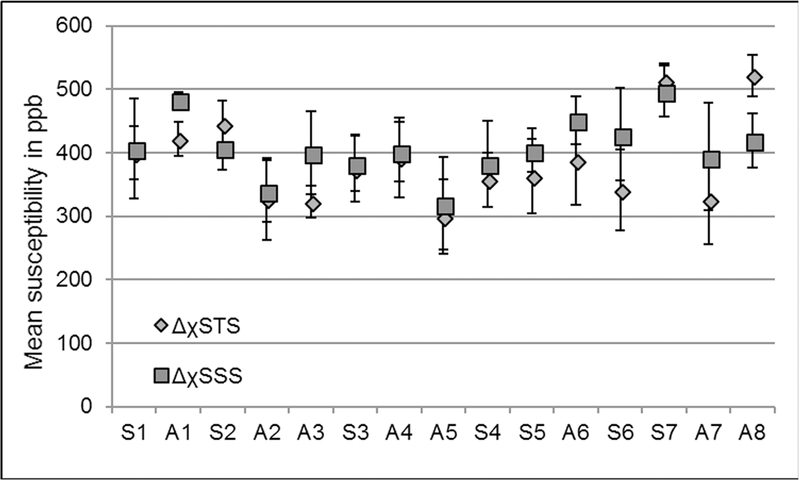
The plot of mean susceptibility values quantified from the dural sinuses from fifteen volunteers. Sn=sagittal and An=axial acquisition, where n is the volunteer number, i.e. n = 1, 2, 3…15. The mean ± σM inside the SSS of all volunteers was 435.3±5.2ppb, where σM represents the variation of the mean between subjects.