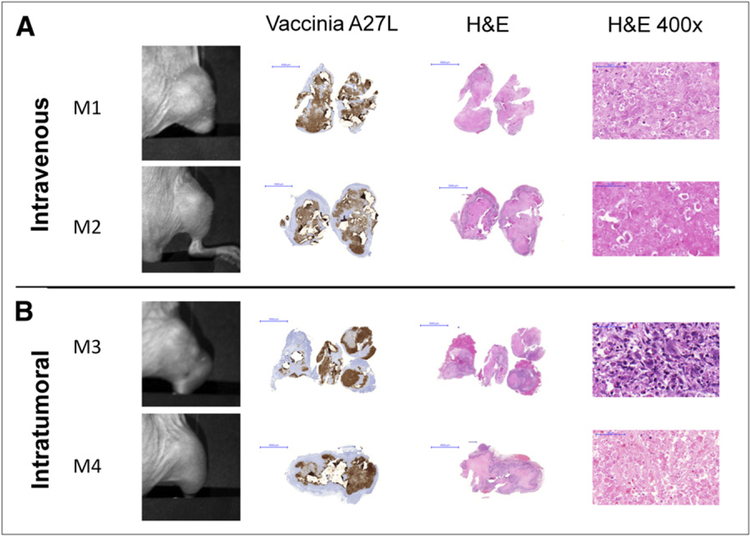
FIGURE 4.
Histologic examination of virus-treated tumors at 5 wk after injection reveals ability of GLV-1h153 to provide insight into tumor biologic activity. (A) Histo-logic examination of imaged tumors at 5 wk after virus injection showed wide areas of necrosis and inflammatory infiltrate. (B) No viable cells were detected histologically in mouse tumors except in mouse 3, which retained some tumor proliferation activity, as seen at ×400 magnification. Presence of GLV-1h153 in tumors was confirmed histologically, shown here with the brownish precipitate against vaccinia A27 L antigen in intravenous and intratumoral groups (A and B, respectively). Bars represent 5,000 μm. M1, M2, M3, and M4 5 mouse 1, mouse 2, mouse 3, and mouse 4, respectively.

