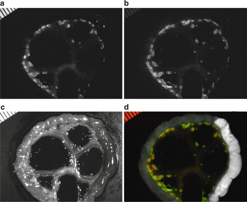
Fig. 1.

Co-staining in a peritoneal-dissemination model of ovarian cancer. Spectral fluorescence images of a SHIN3-RFP tumor-bearing mouse which was injected with GmSA-RhodG. Unmixed spectral fluorescence images display (a) SHIN3-RFP tumors in the RFP spectrum and (b) the tested imaging probe (GmSA-RhodG) in the RhodG spectrum. (c) White light image of gut and mesentery. (d) Overlay of spectral images illustrates the co-localization of both the RFP and RhodG fluorescence.
