
Figure 3.
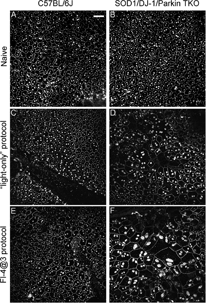
Fundus camera-delivered light induced retinal degeneration leads to more pronounced changes in size and morphology in the RPE cells of SOD1/DJ-1/Parkin-deficient mice (TKO mice) compared to C57BL/6J mice. RPE flat mounts of naive eyes (A,B), eyes treated two weeks earlier with the “light-only” FCD-LIRD protocol (C,D) or the “fluorescein- assisted” (Fl-4@3) FCD-LIRD protocol (E,F) were stained with ZO-1. FCD-LIRD led to increase cell size and increased irregularity in cell shape in both C57BL/6J (A,C,E) and TKO (B,D,F) mice. However, the changes were more prominent in the TKO mice. Scale bar = 50 μm