
Figure 5.
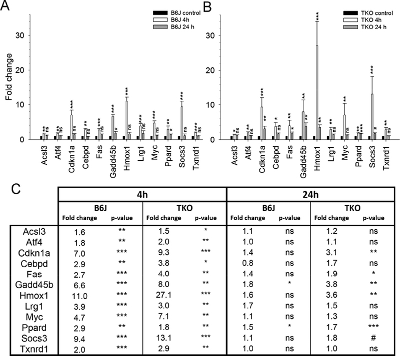
Gene expression analysis using qPCR determined that a large number of oxidative stress-related genes increased after fundus camera-delivered light induced retinal degeneration (FCD-LIRD) in both C57BL/6J (B6J, A) and SOD1/DJ-1/Parkin-deficient (TKO, B) mice. The bars represent gene expression as Fold Change relative to baseline and using GAPDH as an internal control. The expression of most of these genes increased 4h after FCD-LIRD, but moved back towards baseline 24h after treatment (A,B,C). For the analysis, eyes were collected from naive mice (C57BL/6J: n = 8 eyes; SOD1/DJ-1/Parkin-deficient mice: n = 5 eyes), from treated mice 4h after FCD-LIRD (C57BL/6J: n = 5; SOD1/DJ-1/Parkin-deficient mice: n = 4), and from treated mice 24 h after FCD-LIRD (C57BL/6J: n = 4 eyes; SOD1/DJ- 1/Parkin-deficient mice: n = 5 eyes). Statistical analysis is based on a comparison to naive eyes (control). * = p<0.05, ** = p<0.01, *** = p<0.001, ns = no significant difference