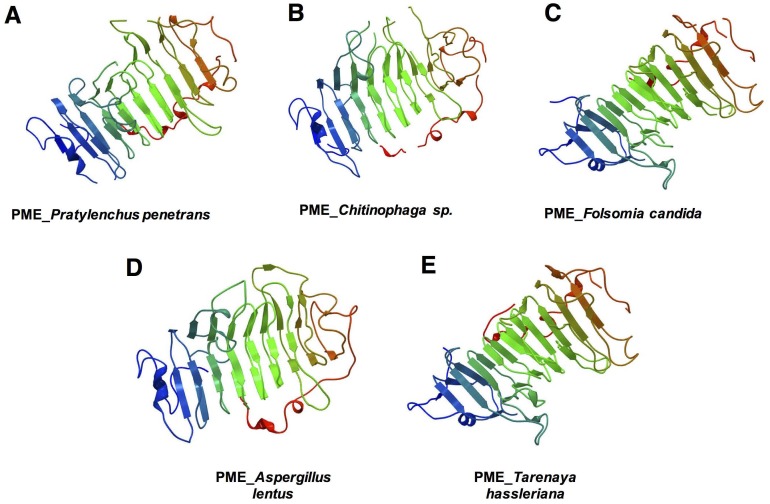
Fig 3. Three-dimensional model predicted for the pectin methylesterase of different organisms.
(A) Pratylenchus penetrans, (B) bacteria (Chitinophaga sp.), (C) collembolan (Folsomia candida), (D) fungi (Aspergillus lentus), and (E) plants (Tarenaya hassleriana). The models of P. penetrans and Chitinophaga sp. were based on the three-dimensional model of the Erwinia chrysanthemi (Phyre2 fold library ID: d1gq8a), F. candida and T. hassleriana were based on the model of Daucus carota (Phyre2 fold library ID: d1qjva), and A. lentus on the model of A. niger (Phyre2 fold library ID: c5c1cA), respectively. The N-terminal is indicated in blue and the C-terminal is shown in red.

