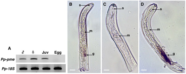
Fig 5. Expression and localization of Pratylenchus penetrans Pp-pme transcripts.
(A) Determination of Pp-pme expression in different nematode developmental stages of P. penetrans by semi-quantitative RT-PCR. As a positive control, all cDNA templates were amplified with primers derived from the 18S gene of P. penetrans. The nematode stages were separated as males, females, juveniles (J2-J4), and eggs. (B-C) Detection of the Pp-pme transcripts by in situ hybridization. Nematode sections were hybridized with antisense (B), or sense (C) Pp-pme digoxigenin-labeled cDNA probes. (D) As a positive control, in situ hybridization was performed with the antisense probe designed for the CWDE (Pp-eng-1) specifically localized within the esophageal glands g: esophageal glands; m: metacorpus; s: stylet.

