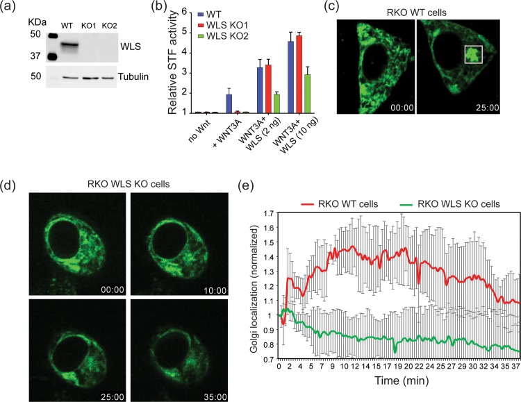
Fig 3. WLS knockout abolishes WNT3A trafficking.
(a) Western blot analysis of WLS-depleted RKO cells. Cell lysates were assessed for WLS protein levels after CRISPR-Cas9 targeting and single cell cloning and tubulin was used as loading control. The blot shows two different CRISPR-Cas9 targeted clones. (b) SuperTopFlash assay in RKO wildtype and RKO WLS KO cells. The assay shows complete loss of Wnt signaling in RKO WLS KO cells and rescue by transfection with 2 ng or 10 ng wildtype WLS expression plasmid. (c) Fluorescence photomicrographs of RKO wildtype cells transfected with RUSH-eGFP-WNT3A illustrate transport from ER (time = 00:00) (minutes:seconds) to Golgi (time = 25:00) after biotin addition. (S4 Video) (d) Fluorescence photomicrographs of RKO WLS KO cells transfected with RUSH-eGFP-WNT3A showing WNT3A does not exit the ER upon biotin addition. (e) Time-dependent analysis of Golgi localization of RUSH-eGFP-WNT3A, analyzed as in Fig 1C and 1D, in wildtype and WLS KO RKO cells. (n = 4 cells per condition ± SD).

