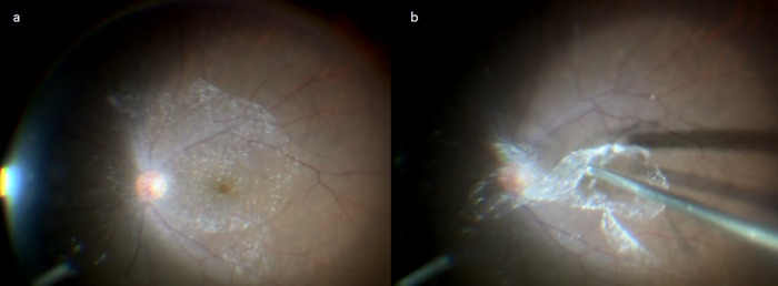
Fig 1. Method used for collection of the bursa premacularis (BPM).
The BPM was visualized with an application of triamcinolone acetonide (TA) to the posterior pole of the vitreous (a). TA accumulated on the anterior surface of the BPM. A diamond-dusted membrane scraper was then used to form a window on a part of the BPM, from which the BPM was separated from the retinal surface by aspiration with a vitreous cutter to be selectively collected (b).

