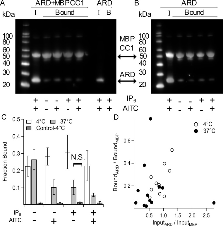
Fig 5. Interaction of MBPCC1 with the N-terminal ARD.
A and B. Anti-His Western blots of amylose-resin purified protein from bacteria expressing both ARD and MBPCC1 or ARD alone. Incubation with resin and subsequent washing of resin was carried out at 4˚C (A) or 37˚C (B). “I” indicates input sample and “Bound” indicates protein pulled down that remained after 4 wash steps. At each temperature lysates were incubated with IP6 and/or AITC. C. Fraction of ARD bound to MBPCC1 normalized to input (see methods). Control indicates amylose purification of cells only expressing ARD at 4˚C. D. Bound ARD/MBP ratio vs Input ARD/MBP ratio showing that more ARD in the input correlates with increased ARD binding to MBPCC1 during purification. Except for the one indicated condition, all differences were significant (p < 0.05).

