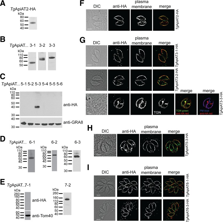
Fig 2. Expression and localization analysis of T. gondii ApiAT family proteins.
(A-E) Western blots with anti-HA antibodies to measure the expression and molecular mass of tagged TgApiAT proteins in tachyzoites stages of the parasite. Western blots with antibodies against GRA8 and Tom40 were used to test for the presence of protein in samples where the HA-tagged TgApiAT protein was not detected. (F-I) Immunofluorescence assays with anti-HA antibodies to determine the localisation of HA-tagged TgApiAT proteins (green in merge). Samples were co-labelled with antibodies against the plasma membrane marker P30 (red in merge). TgApiAT3-3-HA-expressing parasites were co-transfected with the trans-Golgi network (TGN) marker Stx6-GFP [63], and labelled with anti-HA (red in merge), anti-P30 (plasma membrane, PM; blue in merge) and anti-GFP (green in merge) antibodies. All scale bars are 2 μm.

