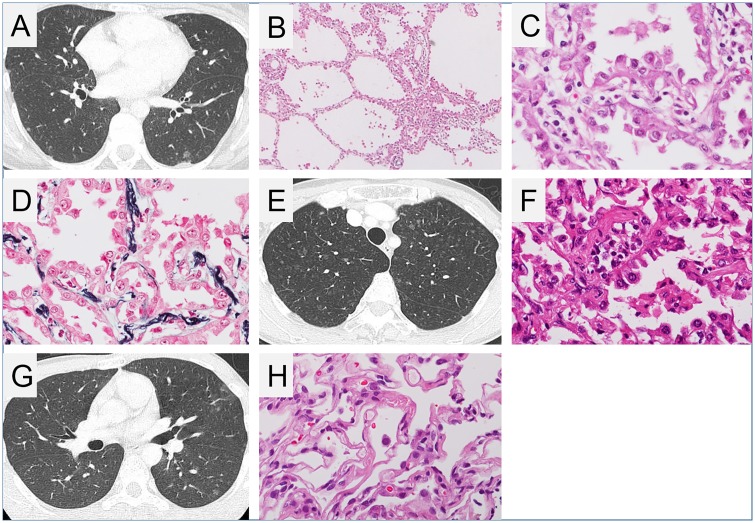
Fig 2. Radiological and histopathological findings of multifocal micronodular pneumocyte hyperplasia.
(A) Chest high-resolution computed tomography (HRCT) image of patient 1 (daughter) showed several ground glass nodules measuring 1–7 mm in size. Cystic changes suggestive of lymphangioleiomyomatosis (LAM) were not observed. (B–D) Histopathological images of multifocal micronodular pneumocyte hyperplasia (MMPH) of patient 1. (B) Low-powered view (×20), haematoxylin and eosin (HE) staining. There were well-demarcated, nodular lesions ranging from 2 to 5 mm in the pulmonary parenchyma. (C) High-powered view (×200), HE staining. Alveolar structure was maintained. Enlarged type II pneumocytes lining the thickened alveolar septa had abundant eosinophilic cytoplasm and spherical nuclei with mild nuclear atypia. (D) High-powered view (×200), Elastica-Masson staining. The alveolar septa were thickened by proliferation of elastic fibres. (E) Chest HRCT image of patient 2 (mother) showed several micro ground glass nodules. (F) Histopathological image of lung specimen of patient 2. High-powered view (×200), HE staining. Multifocal well-demarcated nodular lesions were observed. Enlarged cuboidal type II pneumocytes had swollen nuclei. The alveolar septa were thickened by fibrous proliferation. (G) Chest HRCT image and (H) histopathological image, high-powered view (×200), HE staining, of the lung lesion of patient 3 (son) showed findings consistent with MMPH.

