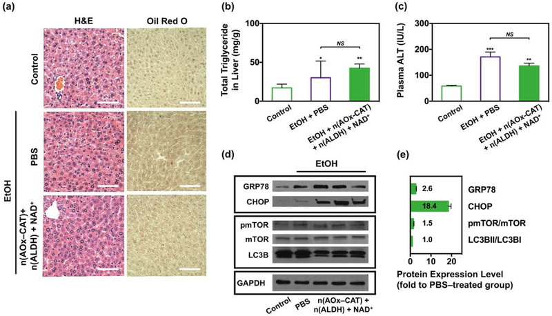
Figure 4.
Biocompatibility of the antidote after HFD and acute alcohol intoxication. (a) Representative H&E and Oil Red O staining of the liver tissues in alcohol–intoxicated mice treated with PBS, or n(AOx–CAT) and n(ALDH) with NAD+ as the antidote. Liver tissue from healthy mice was used as the control. Scale bar, 50 μm. (b) Total liver triglycerides in healthy mice (n=5) and alcohol–intoxicated mice treated with PBS (n=5) or the antidote (n=7). (c) Plasma ALT level in healthy mice (n=5) and alcohol–intoxicated mice treated with PBS (n=5) or the antidote (n=7). (d) Protein expression levels of the ER stress markers (GRP78, CHOP), and autophagy markers including the mechanistic target of rapamycin (mTOR), phosphorylated mTOR (pmTOR) and microtubule–associated protein 1A/1B–light chain 3 (LC3B). (e) Quantification of protein expression levels of the ER stress and autophagy markers, normalized with glyceraldehyde–3–phosphate dehydrogenase (GAPDH). Data are presented as mean ± SEM (n=5~7).

